What Is Hip Impingement?
The hip joint is a ball and socket joint. It consists of the end of your femur (upper leg bone) which has a round end (shaped like a ball) that sits in a cup-shaped socket of your pelvis known as the acetabulum. The acetabulum and femurs both have articular cartilage on their surface to improve joint interaction. At the edge of the acetabulum is another type of cartilage that forms the labrum. The labrum creates a deeper socket and a vacuum-like seal to help keep the ball centered in the socket. On top of that you have a joint capsule that contains the joint, various ligaments that connect the femur and pelvis, and finally muscles that dynamically provide support and function to the hip and pelvis.


A common cause of hip pain in individuals is hip impingement. This is when the ball and socket of the hip joint bump into each other, and it occurs due to faulty mechanics with repetitive movements. This can cause wear and tear on the joint surfaces, potentially contributing to osteoarthritis; as well as increased stress on soft tissue structures of the hip, such as the labrum; causing pain, mobility limitations, muscle inhibition, and further wear and tear on the joint.
The majority of labral tears are associated with having some form of hip impingement, but not all hip impingements have labral tears. With either, the onset of pain can be gradual or with a specific movement. When the onset has occurred with a specific episode, it is often due to a buildup of repetitive movements and the specific episode was “the straw that broke the camel's back.” If the labrum is torn, the hip joint loses some of its stability which creates further wear and tear on the joint surface and requires even more focus on hip mechanics and stability.
What Does Hip Impingement Feel Like? What Are Hip Impingement Symptoms?
Most often, hip impingement will be associated with complaints of “pinching” or pain in the front of the hip at its crease and/or groin. This occurs particularly when the hip is taken into flexion, whether that’s bringing your knee up toward your chest or going into the deeper position of a lunge or a squat, as well as when taking your leg across your body or pivoting and turning over your foot, moving the hip into adduction or internal rotation. Front side hip impingement is most common and associated with more front side hip and groin discomfort, and posterior hip impingement is more likely to cause deep butt pain with hip extension. Occasionally there will be accompanying pain of deep butt discomfort or aching, and this can also be associated with other muscular complaints of greater trochanteric bursitis (pain on the side of the hip) due to compensatory patterns. Pain is often reproduced by taking a very large step.
In symptoms that are further along and possibly associated with a labral tear, you may experience a catching or locking sensation in your hip, particularly after prolonged sitting or walking, or a feeling of instability. If there is further wear and tear progressing to hip osteoarthritis, there will be restrictions in range of motion as well as a firmer end feel.
Hip Impingement Test
In a clinic setting, a clinician will perform a test for hip impingement known as FADIR (flexion, adduction, internal rotation). The patient will lay on their back and the clinician will passively lift the leg up to 90 degrees with their knee bent to take the knee across their body as they also take their foot out to the side. If the patient reports pain or discomfort in the front of the hip, groin, or front side of the hip with the movement, then the test is positive and the patient is deemed to have impingement. This test does not delineate between impingement or a labral tear.
X-ray imaging is used to assess for bony deformities (such as a cam or pincer lesion) and further studies including a CT scan, MRI or preferably MR arthrogram (where dye is injected into the joint) can be used to assess other soft tissue damage (such as in the labrum). A cam lesion is where the ball of the femur is not smooth, but has bumps, is larger, or has a non-cylindrical shape preventing it from moving well in the socket. A pincer lesion is where the roof of the acetabulum extends beyond its normal position, causing the femur to bump into it. People can sometimes have both at the same time.


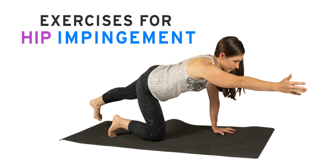
Exercises for Hip Impingement
Free PDF Guide
📑 PDF Guide | 🕓 24 Pages
Exercises for Hip Impingement: Restore Hip Mechanics, Stability & Function
Conditions that can contribute to changes in the femur and acetabulum are genetics; repetitive exposure to sports with a high level of hip flexion and rotation like soccer, football, hockey, gymnastics, and dance during adolescence that can trigger adaptive remodeling; and childhood hip disease such as Legg-Calve-Perthes, slipped capital femoral epiphysis, and coxa vara. Legg-Calve-Perthes disease is where part of the hip joint doesn’t get enough blood, causing the bone to die and affecting the articulation in the joint. Slipped capital femoral epiphysis is a separation of the ball from the thigh bone at the growth plate in adolescents, and is more common in obsese children. Coxa vara is a less common condition where the thigh bone and ball do not grow at the same rate, which changes the angle that the femur enters into the socket.
All that being said, research showed that in asymptomatic individuals (those without symptoms or pain) 73% were found to have bony abnormalities, like a cam or pincer lesion, and 69% were found to have labral tears. https://pubmed.ncbi.nlm.nih.gov/23104610/. Anecdotally, it is also common for individuals to have similar findings on their symptomatic leg as their non-symptomatic leg, so I pose the question: Why is one leg having pain and the other isn’t, if they both look similar? Imaging should be used as a piece to the puzzle, with another important piece being movement assessment. The positive findings in imaging could lead to less room for error in your movement patterns, but it doesn’t mean you can’t overcome it or must fall victim to the diagnosis.
Another form of assessment as well as a treatment used by doctors is to inject the hip with a corticosteroid and local anesthetic under the use of fluoroscopy (a special type of live X-ray) to help guide the needle directionally into the joint. Some doctors will perform more topical injections at bursa sites that involve addressing more of the inflammation of the soft tissue, but these do not get directly into the joint and assess the root cause. For this, the injection needs to be done with the use of fluoroscopy. If a patient has a change in symptoms after the injection, this is noted as a positive finding that the joint irritation was the cause of the pain. Some doctors will end it there, instructing the patient to return to activities of daily living and come back for further treatment if needed, when in reality the patient should take advantage of not being in the acute stage of inflammation and address the muscle imbalances that contributed to the pain to begin with.
Hip Impingement Causes
Bony anatomy changes in the femoral head or acetabulum as well as increased hip instability created by labral tears can contribute to hip impingement, but muscles dictate how bones move and joints interact. It’s therefore important to look at hip and pelvis alignment and muscle function.
Often, impingement will occur due to a pelvis that is in more of a tipped forward, anterior pelvic tilt position. Labral tears can occur due to constant pinching of the labrum or can be associated with a femur that is positioned further forward in the socket due compensatory patterns.
In proper hip mechanics, as the leg comes up into flexion, the ball of the femur will glide back into the socket. On the pelvic side, if a pelvis is in more of a tipped forward posture at rest, then the femur is already in some relative hip flexion with the roof of the acetabulum already covering over the femur, so the hip will “run out of room” to go into deeper flexion. The pelvis being in more of a tipped forward posture also orients the femurs in an externally rotated and abducted position (think turned out) which will then limit internal rotation and adduction due to the bony block at the pelvis, causing pain or discomfort with that movement. So instead of focusing on femur range of motion, it’s often best to address pelvic positioning first and then approach femur position.
Dynamically, being able to eccentrically lengthen the glutes and stabilize the pelvis will allow for this ball to move in the socket. If the back side is compressed, then you’ll either have a pelvis that tips forward, lengthening from the hamstrings instead and bringing the roof down, or an overly tucked pelvis that doesn’t create room for the femur to glide back, putting pressure on the front. Lack of lengthening in the back side of the pelvis will also affect pelvic position such that the sockets will be oriented where the femurs are biased in the turned out position. This will further contribute to hip imbalances with back side tightness and limited hip internal rotation and adduction.
Hip Impingement Treatment
Some hip impingement will require surgery, but the first line of approach should always be conservative through movement assessment and exercise. Surgery is most commonly performed arthroscopically, where 3-4 small incisions are done to access the joint with the use of internal cameras to assist in the procedure. This surgery can also be used as a diagnostic tool in an exploratory procedure. The recovery from surgery can depend on the severity of the bony changes and presence of a labral tear.
That being said, even if you end up being a candidate for surgery, addressing the muscle imbalances as much as you can first will help set you up for that much more success afterwards. With surgery, you’ll have to work through the acute changes it causes where you’re often non or partial weight bearing with range of motion restrictions, and basically “lost a knife fight” you have to recover from.
Hip Impingement Surgery
A typical recovery after arthroscopic hip surgery can vary depending on various factors, from tissue quality to the number of procedures performed (cam, pincer, or both being shaved down, labral repair versus debridement, bursectomy, or microfracture to improve cartilage) to nutrition and past medical history. With someone who has only hip impingement, the lesion is shaved down to improve the fit of the ball in the socket. Someone who had just debridement and shaving of the bone is often on more of an “as tolerated” protocol without worrying about protecting repaired tissue. Often they will have crutches for 1-2 weeks and return to full physical activity after three to 6 months. That being said, some of these types of procedures can be “salvage” procedures where the patient is being bought some time before needing a full hip replacement.
However, if other procedures occur, such as a labral repair where the labrum is sewn back together and not just cleaned up, it’s important to avoid putting stress on the repaired tissue during the initial healing time frame. This means a longer recovery with limited range of motion and weight bearing during the beginning stages. This is important for the tissue to heal, and it takes priority; but it can also lead to other soft tissue restrictions and overload that will have to be overcome as the individual heals from the surgery. Doctors will have varied protocols of weight bearing limitations ranging from 2 to 8 weeks, range of motion restrictions for the first month, other activity restrictions, and some will use hip braces and others not. Full recovery can take 6 months to a year depending on the surgery and activity level that is desired.
It’s great that surgery has come a long way with what can be done through small incisions, but it can be hard to realize how much was addressed internally and therefore how much needs to heal. Seeking the assistance of an in-person physical therapist who is familiar with hip surgeries is an important part of optimal recovery.
Be sure to check out part 2 of this article where we go over hip impingement exercises you should work on if you are trying to avoid surgery!

Free Pelvis Webinar (Professionals)
Understanding Deep Hip Rotator Tightness
🎥 1 Video Lesson | 🕓 47 Min
Identify gripping patterns, explore proximal hamstring activation, and try practical exercises for mobility and control.
Assess & correct deep hip rotator tightness with targeted tests, hamstring activation, and advanced hip control drills to reduce guarding and improve full hip motion.